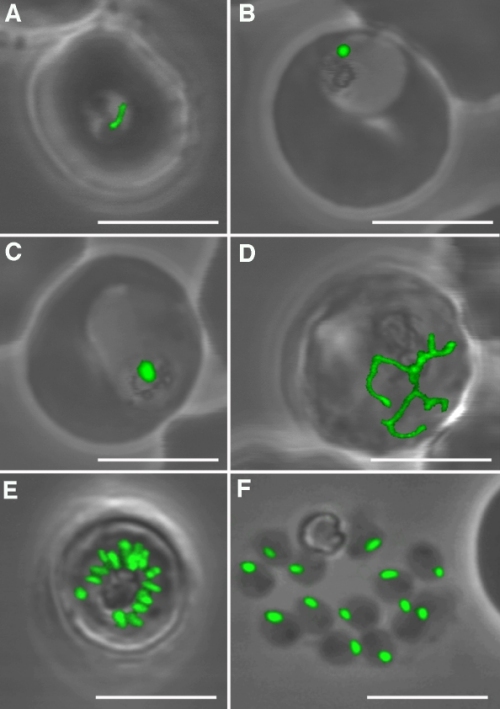
Figure 2. Plasmodium falciparum with its chloroplast-related organelle, the apicoplast. Here the morphology of the apicoplast is shown throughout the asexual life cycle of the blood-stage plasmodium parasites through the use of green fluorescent protein (GFP). The single apicoplast in A enlarges and elongates to form the branched forms shown in D. They divide (E) and segregate into the free merozoites in F. Source: Figure 5 from Waller R.F., Reed M.B., Cowman A.F., and McFadden G.I. 2000. Protein trafficking to the plastid of Plasmodium falciparum is via the secretory pathway. EMBO J. 19: 1994–1802. http://www.nature.com