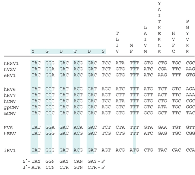
Figure 27.2. Designing PCR primers. Alignment of a region of the DNA polymerase genes from a sample of herpesviruses (abbreviations of the virus names given in leftmost column). The different amino acids present at each position in these samples (e.g., the first amino acid is Y in all of the viruses, the seventh varies among TLIV) are shown above the top horizontal bar. An alignment of the DNA sequences encoding these proteins is shown with conserved nucleotides highlighted in blue and with each codon separated. Note that even related viruses frequently use different codons for the same amino acid. “Degenerate” PCR primers could be designed from this alignment that take into account the variation in codon usage and even the choice of amino acid. Such primers would include a mixture of all the possible sequences. Even when a protein’s amino acid sequence is 100% conserved between species, the degeneracy of the genetic code usually prevents the use of nondegenerate primers. This is an important consideration because PCR works better with less degenerate primers. (Modified from Rose T.M. Nucleic Acids Res. 26: 1628–1635, Fig. 2, © 1998 Oxford University Press.)
| © 2007-2010 by Cold Spring Harbor Laboratory Press. All rights reserved. |
| The reproduction, modification, storage in a retrieval system, or retransmission, in any form or by any means, electronic, mechanical, or otherwise, for reasons other than personal, noncommercial use is strictly prohibited without prior written permission. You are authorized to download one copy of the material on this Web site for personal, noncommercial use only. The material made available on this Web site is protected by United States copyright laws and is provided solely for the use of instructors in teaching their courses and assessing student learning. Dissemination or sale of any of this material, as a whole or in parts (including on the World Wide Web), is not permitted. All users of these materials and visitors to this Web site are expected to abide by these restrictions. Requests for permission for other uses of this material should be directed to Cold Spring Harbor Laboratory Press, 1 Bungtown Road, Cold Spring Harbor, NY 11724 or submitted via our World Wide Web Site at http://www.cshlpress.com/. |