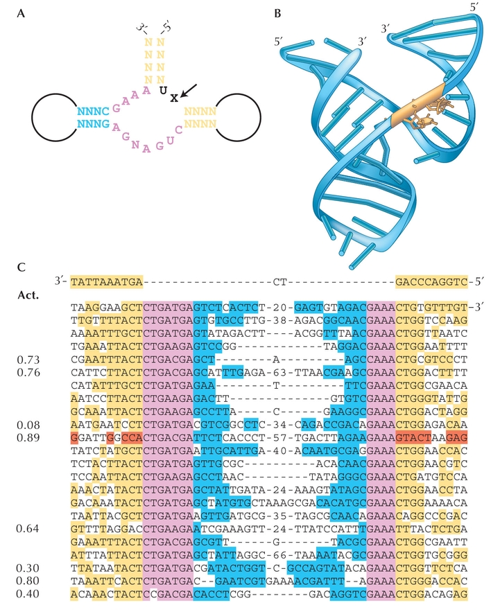
Figure WN20.3. De novo evolution of the hammerhead ribozyme. (A) The secondary structure consists of three double helices. The arrow shows the site at which the hammerhead catalyzes its own cleavage. The core sequence at the catalytic site is shown in magenta; yellow and blue show stem regions that base-pair. X is A, U, or C. (B) The three-dimensional structure, with the cleavage site in gold. (C) Alignment of sequences present after 12 generations. Colors show similarity to the different regions of the natural hammerhead ribozyme; red shows one clone with a variant of the natural stem structure. The blue, yellow, and red sequences are diverse, but do show the base pairing required to maintain the hammerhead secondary structure (A,B). The catalytic activity is shown and is comparable with the natural ribozyme (~0.8 per minute). (These are DNA sequences: T corresponds to U in the RNA sequence.) (A,C, Redrawn from Fig. 3a,b in Salehi-Ashtiani and Szostak 2001. B, Redrawn from Fig. 1b in Doudna and Cech 2002.)
| © 2007-2010 by Cold Spring Harbor Laboratory Press. All rights reserved. |
| The reproduction, modification, storage in a retrieval system, or retransmission, in any form or by any means, electronic, mechanical, or otherwise, for reasons other than personal, noncommercial use is strictly prohibited without prior written permission. You are authorized to download one copy of the material on this Web site for personal, noncommercial use only. The material made available on this Web site is protected by United States copyright laws and is provided solely for the use of instructors in teaching their courses and assessing student learning. Dissemination or sale of any of this material, as a whole or in parts (including on the World Wide Web), is not permitted. All users of these materials and visitors to this Web site are expected to abide by these restrictions. Requests for permission for other uses of this material should be directed to Cold Spring Harbor Laboratory Press, 1 Bungtown Road, Cold Spring Harbor, NY 11724 or submitted via our World Wide Web Site at http://www.cshlpress.com/. |